A small bowel magnetic resonance imaging (MRI) scan is an imaging procedure used to look at the small intestine (bowel) for inflammation, swollen lymph nodes, thickening or any other changes of the bowel wall. These changes could indicate Crohn’s disease.
It is a common procedure for those with inflammatory bowel disease (IBD) or suspected IBD.
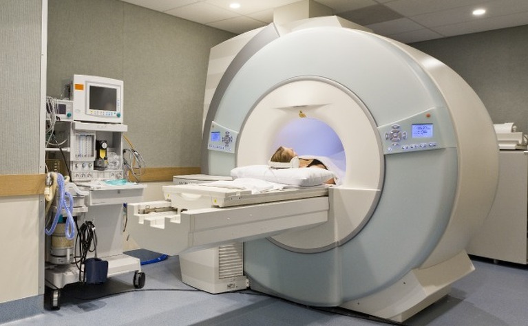
MRIs are a way of generating a 3D image using a combination of powerful magnets and radio waves.
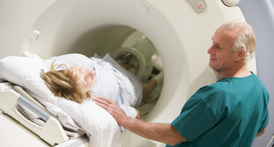
The big cylindrical tunnel you lie in is actually a very powerful magnet, which is used to obtain images of a target body part (in this case, the length of your small bowel).
Your body tissues contain water molecules which are comprised of hydrogen and oxygen ions. MRI scanners magnetise the protons at the centre of hydrogen ions, pulling them into alignment.
Then, short bursts of radio waves are used to knock the protons out of alignment. When these bursts are turned off, the protons in the water molecules line up again, which sends out a signal that is picked up by the machine.
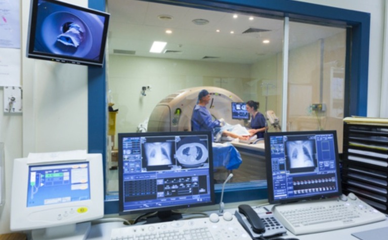
This means that different types of body tissue (e.g. fat vs. muscle) emit different signals due to the speed of the protons realigning, which allows the radiography team to generate an image of the bowel based on the magnetic resonances picked up by the machine.
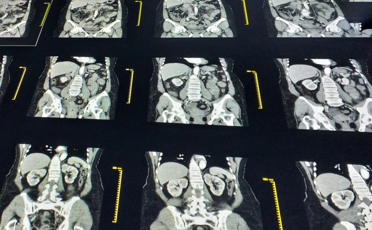
The information obtained from the signals can be used to produce a 3D representation of the body tissues, which radiologists use to identify inflammation or irregular thickening in the bowel.
There are multiple reasons you might be sent for a small bowel MRI scan. The five main reasons for a small bowel MRI are:
When you are invited for a small bowel MRI, this will often be to make sense of symptoms or results from blood tests or stool tests. Together with the results from other tests, an MRI can inform a diagnosis of IBD.
If you are experiencing new symptoms, such as nausea, vomiting, or weight loss, this can indicate a narrowing of the small bowel (such as a stricture). An MRI can help to identify strictures and reveal whether the stricture is the result of active inflammation or scar tissue.
New symptoms can also be the result of abnormal connections between different areas of bowel (fistulae). An MRI scan is the most effective way to determine which of the possible causes is responsible for the change in symptoms.
After an operation or new course of medication for your IBD, your doctor may want to check the treatment is having the intended effect on your Crohn's disease or ulcerative colitis by monitoring inflammation in your bowel.
Sometimes, doctors will use a procedure known as video capsule endoscopy or VCE (whereby a capsule containing a camera is swallowed to take photographs of the lining of the bowel. This is to look for ulcers and inflammation). A small bowel MRI may be conducted prior to the VCE to screen for any strictures or narrowings, because any narrowing in the bowel may cause the camera to get stuck. A small bowel MRI is therefore a useful way of ensuring the safety of the video capsule endoscopy procedure.
As with the VCE, sometimes a small bowel MRI will be carried out prior to surgical procedures on the bowel to check for any other areas of inflammation that may need surgery or other treatment.
An MRI has several advantages to alternative procedures.
Colonoscopy is an intrusive procedure, and while it is more sensitive than an MRI scan in detecting IBD, it only gives access to the large bowel and the end of the small bowel.
Meanwhile, ultrasound is the safest and most comfortable option, but it doesn’t provide information about the full length of the bowel.
MRI gives a more comprehensive view of the bowel, with a good degree of sensitivity and accuracy.
Sometimes you may need to have several different procedures or tests. The results from all of these will help form a picture of what is going on inside your body and help with a diagnosis of IBD or to monitor whether treatment is working.
Generally speaking, an MRI scan is considered to be a very safe procedure. However, there are some possible complications for patients with metal implants and pacemakers. Some also experience side effects caused by the MRI contrast medium which you may need to take for your small bowel MRI scan.