Scans and X-rays for inflammatory bowel disease (IBD)
There are various types of scans and X-rays which are used to both help diagnose inflammatory bowel disease (IBD) and also to monitor it. The results of the scans will be used alongside other tests to build a picture of your IBD.
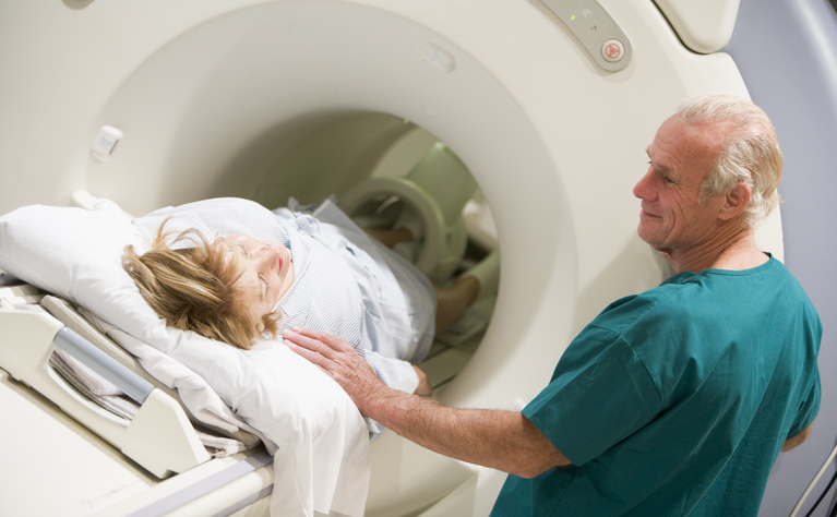
Computerised tomography (CT) (also known as CAT scan) or computerised tomography enterography/enteroclysis (CTE) scans. Several X-rays of your small intestines are taken and assembled by computer to create a detailed image
Ultrasound is used to help diagnose perianal Crohn’s disease as well as examine organs in the abdomen (such as liver and gallbladder). It may also be used to examine the small bowel for blockages
Leukocyte scintigraphy (white blood cell scans)
Magnetic resonance enterography/enteroclysis (MRE) - magnetic fields and radio waves are used to produce detailed images of your small intestines