

Get IBD info delivered to your inbox
Sign up to our mailing list and receive regular articles and tips about IBD.
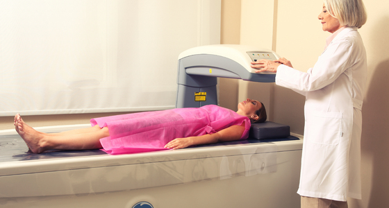
A DEXA scan is a special type of X-ray that measures bone mineral density (BMD).
DEXA stands for "dual energy X-ray absorptiometry". This type of scan is also often known as DXA, or "dual X-ray absorptiometry". It's also sometimes referred to as a bone density scan or a bone densitometry scan.
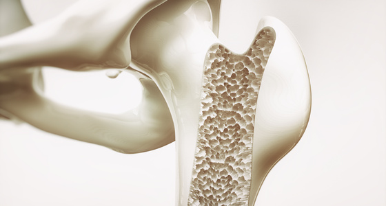
DEXA scans are often used to diagnose osteoporosis (when the bones become weak and fragile, and are more likely to break).
They can also be used to assess the risk of osteoporosis developing in women aged over 50 and in men over 60.
As well as being quick and painless, a DEXA scan is more effective than normal X-rays in identifying low bone mineral density.
If you're over 50 years of age, you may need to have a DEXA scan if you're at risk of developing osteoporosis. If you're under 50, you may need a DEXA scan if there are other risk factors at play, such as smoking or if you have had a previous fracture.
Such risk factors are used in the World Health Organization (WHO) 10-year Fracture Risk Assessment Tool, which applies to both men and women between 40 and 90 years of age.
The tool can be used to assess if a DEXA scan is appropriate, and also uses what is known as the DEXA femoral neck bone mineral density T score to calculate the risk of fracture.
Osteoporosis can affect people of both sexes and all ages, although older, post-menopausal women are particularly at risk. This is because after the menopause the level of oestrogen declines, resulting in a decrease in bone density.
The denser your bones, the stronger and less likely they are to fracture (break). Osteoporosis doesn't cause any symptoms until a bone is broken. It used to be difficult to measure bone density and identify those at risk of developing osteoporosis until a fracture occurred.
However, by using bone densitometry techniques such as DEXA scans, it's now possible to measure bone density before someone gets a fracture.
Read more about when DEXA scans are used.
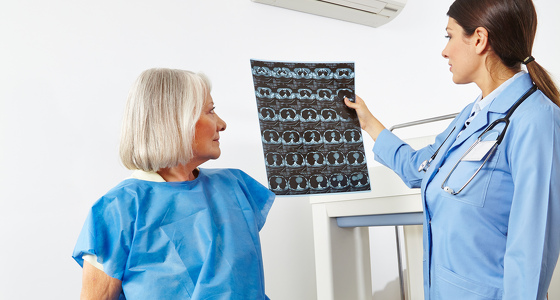
During a DEXA scan, X-rays will be passed through your body. Some radiation will be absorbed by the bone and soft tissue, and some will travel through your body.
Special detectors in the DEXA scanner measure how much radiation passes through your bones, and this information is sent to a computer.
Your bone density measurements will be compared with the bone density of a young healthy adult or an adult of your own age, gender and ethnicity.
Read more about how DEXA scans are carried out.
DEXA scans use a much lower level of radiation than standard X-ray examinations, which means that the radiographer (the technical specialist carrying out the scan) can stay in the scanning room with you during the scan.
The amount of radiation used during a DEXA scan will vary depending on the area of the body being examined, but is very low and less than two days' exposure to natural background radiation (NBR).
By comparison, a chest X-ray uses the equivalent of about five days' exposure to NBR, and a flight to North America is equivalent to about a week's exposure to NBR.
Despite being very safe procedures, DEXA scans and X-rays aren't recommended for pregnant women, as X-rays can damage an unborn child.
Read more about your health during pregnancy.

Why not sign up to our mailing list and receive regular articles and tips about IBD to your inbox?